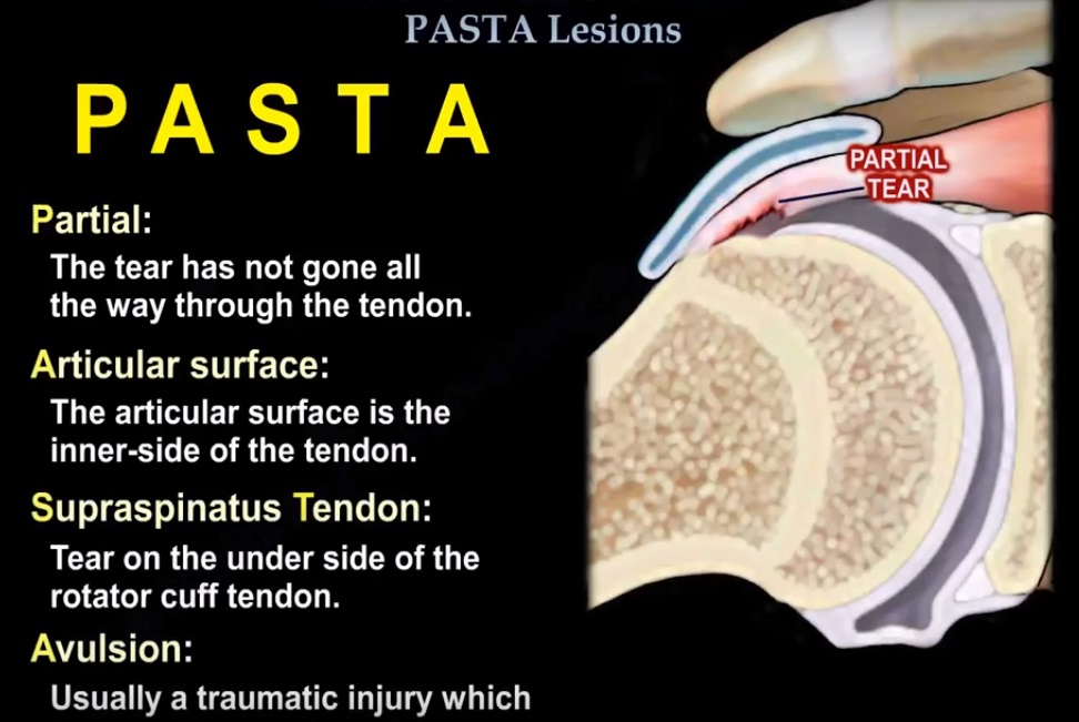
Courtesy: Prof Nabil Ebraheim, University of Toledo, Ohio, USA
- PASTA lesions are usually seen in athletes.
- The tear of the rotator cuff is partial and on the articular surface side.
- There is supraspinatus tendon avulsion and it is difficult to diagnose.
- An arthrogram may help in the diagnosis.
- The tear could be seen on an ultrasound or MRI.
- The MRI arthrogram is done in the ABER position (abduction/external rotation) and is more accurate in showing this lesion.
- The arm will be above the head in the scanner. The normal rotator cuff is about 10-12 mm in thickness.
- If exposed bone between the rotator cuff and the articular margin is more than 7 mm, then there is an at least 50% thickness tear. This is a classic indication for surgery.
- When the lesion is less than 50% and painful, you can debride it. If the lesion is more than 50% and painful, you can repair it.
- The physician may complete the tear to become a full thickness tear, then repair the tear.
- Rotator tears can be full thickness tears or partial thickness tears. PASTA tears may be associated with internal impingement, which is different than external impingement.
- With external impingement, it is subacromial impingement (bursal pathology). In internal impingement, the pathology is on the undersurface of the cuff, so PASTA tears may be associated with internal impingement.

Leave a Reply