ANATOMY OF PERONEUS LONGUS MUSCLE
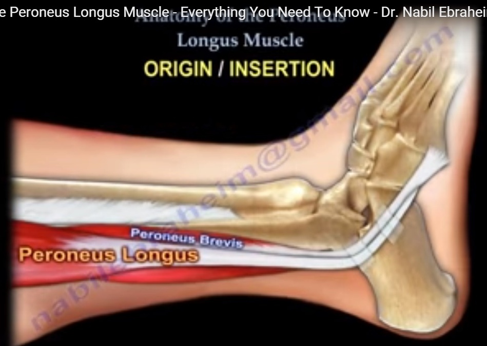
- The peroneus longus muscle passes down the outside of the lower leg and on top of the peroneus brevis.
- The peroneus longus is a longer muscle and has a larger muscle belly compared to the peroneus brevis.
- Peroneal Longus muscle is located within the LATERAL compartment of the lower leg.
- The LATERAL compartment of the lower leg contains both the peroneus longus and the peroneus brevis muscles.
- The LATERAL compartment also contains the superficial peroneal nerve. Both muscles of the lateral compartment (peroneus longus & brevis) are innervated by the Superficial Peroneal nerve.
ORIGIN / INSERTION
- Both the peroneus longus and the peroneus brevis muscles originate from the fibula shaft of the fibula.
- The origin of the peroneus longus muscle comes from the head of the fibula and the upper 2/3 of lateral aspect of the fibular shaft.
- It also originates from the anterior and posterior intermuscular septa of leg.
- The peroneus longus muscle passes over the peroneus brevis muscle.
- The peroneus longus tendon is inserted into the plantar posterolateral aspect of the medial cuneiform and lateral side of 1st metatarsal base.
- The peroneus brevis tendon runs distally and inserts into the 5th metatarsal base.
The peroneus longus tendon is inserted into the plantar posterolateral aspect of the medial cuneiform and lateral side of 1st metatarsal base.
There are two peroneal retinacula which hold the two peroneal tendons at the ankle (superior & inferior).
- The superior peroneal retinacula is more important than the inferior retinaculum. The superior retinaculum is located in the distal 3 cm of the fibula.
- The superior retinaculum originates from the posterolateral ridge of the fibula and inserts into the lateral calcaneus (prevents subluxation).
- Behind the fibula, the tendons run in a sulcus called the peroneal groove which is found on the fibula posteriorly.
- The tendons are stabilized by the superior peroneal retinaculum and the cartilaginous rim.
- Within the groove, the peroneus brevis tendon is anterior and medial to the peroneus longus tendon. Both tendons curve anteriorly around the the tip of the fibula.
- The peroneal tendons are contained in a common synovial sheath.
- Both tendons curve around the tip of the fibula anteriorly with the peroneal tubercle separating the tendons at the level of the calcaneus.
- At the level of the tubercle, the peroneus brevis is dorsal and the peroneus longus is plantar.
- The peroneus longus tendon curves sharply, making a 90° turn medially and passing in a groove beneath the cuboid where it crosses to the plantar aspect of the foot before inserting medially into the base of the 1st metatarsal and the medial cuneiform.
Innervation
- The superficial peroneal nerve supplies the peroneal muscles (L5, S1, S2).
Function
- The peroneus longus muscle everts the hindfoot, flexes the ankle.
- The peroneus longus also plantar flexes the first ray (very important function).
SOME CONDITIONS THAT AFFECT THE PERONEUS LONGUS MUSCLE & TENDON
1. Peroneus Longus Tendonitis (inflammation)
2. Peroneus Longus Tendon Rupture or Tear-Tears of the peroneus longus tendon are not common and usually occur at the peroneal tubercle. Tears of the peroneus brevis tendon usually occur at the level of the peroneal groove.
3. Os Peroneum: There is an interesting observation involving rupture of the peroneus longus tendon that needs to be explained.
- Os peroneum is a sesamoid bone present within peroneus longus tendon in about 20% of people’s feet and is normally located less than 1cm proximal or distal to calcaneocuboid joint.
- It should not be confused with Os Vesalianum pedis accessory bone or an apophysis of fifth metatarsal.
- The oblique course of the peroneus longus tendon within the cuboid groove and the presence of the Os Peroneum along with sudden movement such as inversion and supination of the foot may cause rupture of the tendon at the site of the ossicle.
- If the Os Peroneum has proximal migration more than 1 cm from the calcaneocuboid joint, rupture of the peroneus longus tendon is likely.
- The Os Peroneum is bilateral in about 60% of patients and the ossicle is bipartite in 30% of patients. Fracture of Os peroneum is rare.
- Fragment seperation of 6 mm or more is associated with rupture of the peroneus longus tendon.
- 4. Subluxation or dislocation of Peroneus longus tendon
The patient may feel a POP or a SNAP sensation as the peroneal tendons move over the lateral malleolus due to rupture of the superior peroneal retinaculum.
Courtesy: Prof Nabile Ebraheim, University of Toledo, Ohio, USA
nice