Courtesy: Susan Mackinnon, Andrew Yee, University of Washington School of Medicine, St Louis, USA
Posterior Interosseous Nerve Release
Standard Edition (1.130823.130731)
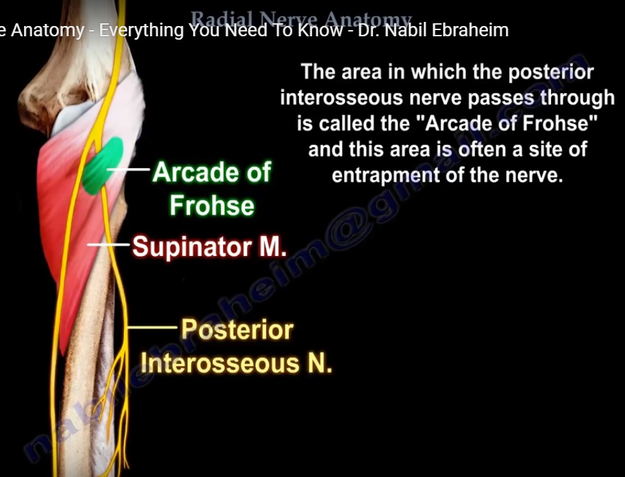
Compression of the posterior interosseous nerve can exhibit clinical weakness or functional loss of finger / thumb extension and lack of ulnar wrist extension. Provocative tests can confirm whether the radial tunnel in the region of the posterior interosseous nerve is the area of compression. Multiple anatomical structures can be involved in compressing the nerve in the tunnel, however the primary site of compression is the arcade of Froshe; this being the tendinous proximal border of the superficial head of the supinator. Other compressive structures can include the radial recurrent vessels (Leash of Henry) and tendinous proximal border of the extensor carpi radialis brevis. Decompression of the posterior interosseous nerve involves releasing these structures. If there is an associated lateral epicondylitis, release of the extensor carpi radialis brevis is taken further laterally. In this case, this patient presented with a recovering traumatic C7,8,T1 plexus injury. However, recovery of radial nerve function was halted for a few weeks with marked discomfort over the radial tunnel. Release of the posterior interosseous nerve was elected to promote more prompt and complete functional recovery.
Table of Contents (Standard)
00:20 Orientation
00:25 Incision / Exposure
00:54 Identifying the Posterior Antebrachial Cutaneous Nerve
01:26 Identifying the Interval between the Brachioradialis and ECRL
01:35 Exposing the Interval and Plane between the Brachioradialis and ECRL
02:09 Identifying the Superficial Branch of Radial Nerve
02:29 Identifying the Radial Recurrent Vessels and ECRB Nerve
02:36 Ligating the Radial Recurrent Vessels
02:54 Identifying the Posterior Interosseous Nerve
03:05 Identifying / Releasing the Tendinous Proximal Border of ECRB
04:25 Identifying / Releasing the Tendinous Proximal Border of Supinator
05:33 Releasing Distal Tendinous Fibers of Supinator
05:46 Proximal Exposure for Additional Compressive Structures
06:01 Radial Nerve Anatomy in the Forearm

excellent explanation….
Excellent informative video with candid explanation .